Key Points
Type-1 diabetics exhibit lower resting oxygen saturation, lower cardiovascular control, reduced hypoxic chemoreflexes, and enhanced hypercapnic chemoreflexes
The root cause of these problems is resting tissue hypoxia, which causes over-activation of the sympathetic nervous system and autonomic and cardiovascular dysfunction
Autonomic imbalance in diabetes is largely functional, and therefore reversible
The Breathing Diabetic Summary
This is a follow-on to our previous paper on cardio-respiratory control in diabetes. This paper, however, is a clinical study rather than a literature review.
Previous studies have shown respiratory problems in diabetics. Previous studies also have shown cardiovascular dysfunction in diabetics. However, no studies simultaneously examined both of these factors in an integrated fashion. Thus, the aim of this study was to comprehensively examine cardio-respiratory function in type-1 diabetics.
The key measurements from this paper were resting oxygen saturation, baroreflex sensitivity (BRS; a marker of cardiovascular and autonomic control), and both hypoxic and hypercapnic chemoreflexes (markers of respiratory control).
Their hypothesis: If the BRS and chemoreflexes were suppressed in diabetics, this would indicate nerve damage was present. However, if cardiovascular function was suppressed, while chemoreflexes were enhanced, this would indicate autonomic imbalance that has a functional cause. In this latter case, therapies aimed at restoring cardio-respiratory control (for example, slow breathing) could help prevent diabetic complications.
The study had 46 patients with type-1 diabetes and 103 age-matched control subjects. The participants went through a variety of tests to evaluate baroreflex functioning and chemoreflexes. For example, to measure the patients’ hypercapnic chemoreflex, oxygen was kept constant while CO2 was gradually increased. The chemoreflex can then be measured as the slope of the relationship between minute ventilation and change in CO2 (or oxygen in the case of the hypoxic chemoreflex). A large change in minute ventilation for a small change in CO2 would represent an enhanced hypercapnic chemoreflex.
Interestingly, the results showed that although diabetics displayed larger breathing volumes than controls, they had slightly higher CO2 levels and reduced oxygen saturation. However, they did have an enhanced hypercapnic chemoreflex, meaning they could not tolerate changes in CO2 as well as controls. And, somewhat surprisingly, they had a reduced hypoxic chemoreflex, meaning they could tolerate lower oxygen levels without increasing their breathing as much as controls.
The diabetics also exhibited a lower resting oxygen saturation. This is fascinating because the lower resting oxygen saturation implies a significantly reduced partial pressure of oxygen (due to the oxyhemoglobin dissociation curve). This would result in tissue hypoxia. What’s more, they cite a paper (which is now near the top of my reading list) that shows that a high HbA1c also reduces tissue oxygenation by increasing oxygen’s affinity to hemoglobin (shifting the dissociation curve to the left).
The authors suggest that their results can be interpreted as follows: Resting tissue hypoxia, combined with a suppressed hypoxic chemoreflex, leads to an enhanced compensatory hypercapnic chemoreflex and chronic activation of the sympathetic nervous system. This, in turn, leads to a suppression of the cardiovascular system (reduced BRS and reduced heart rate variability). It’s a vicious cycle.
However, this is actually great news. Their results suggest that diabetic autonomic imbalance is largely functional and not related to nerve damage. (Remember, both the cardiovascular reflexes and the chemoreflexes would have been suppressed with nerve damage). In fact, the authors suggest that this imbalance likely leads to nerve damage rather than being the result of it. Therefore, therapies targeting cardio-respiratory control could help reverse/prevent diabetic complications.
Finally, the authors suggest that breathing control and physical exercise could be two such therapies to restore cardio-respiratory function. We know that slow breathing has many therapeutic benefits for the cardiovascular, autonomic, and respiratory systems. And, we know that slow, light breathing increases CO2 and increases tissue oxygenation (due to the Bohr effect). Now, we know that these positive benefits have the potential to stop or reverse diabetic complications.
Abstract from Paper
BACKGROUND: Cardiovascular (baroreflex) and respiratory (chemoreflex) control mechanisms were studied separately in diabetes, but their reciprocal interaction (well known for diseases like heart failure) had never been comprehensively assessed. We hypothesized that prevalent autonomic neuropathy would depress both reflexes, whereas prevalent autonomic imbalance through sympathetic activation would depress the baroreflex but enhance the chemoreflexes.
METHODS: In 46 type-1 diabetic subjects (7.0±0.9year duration) and 103 age-matched controls we measured the baroreflex (average of 7 methods), and the chemoreflexes, (hypercapnic: ventilation/carbon dioxide slope during hyperoxic progressive hypercapnia; hypoxic: ventilation/oxygen saturation slope during normocapnic progressive hypoxia). Autonomic dysfunction was evaluated by cardiovascular reflex tests.
RESULTS: Resting oxygen saturation and baroreflex sensitivity were reduced in the diabetic group, whereas the hypercapnic chemoreflex was significantly increased in the entire diabetic group. Despite lower oxygen saturation the hypoxic chemoreflex showed a trend toward a depression in the diabetic group.
CONCLUSION: Cardio-respiratory control imbalance is a common finding in early type 1 diabetes. A reduced sensitivity to hypoxia seems a primary factor leading to reflex sympathetic activation (enhanced hypercapnic chemoreflex and baroreflex depression), hence suggesting a functional origin of cardio-respiratory control imbalance in initial diabetes.
Journal Reference:
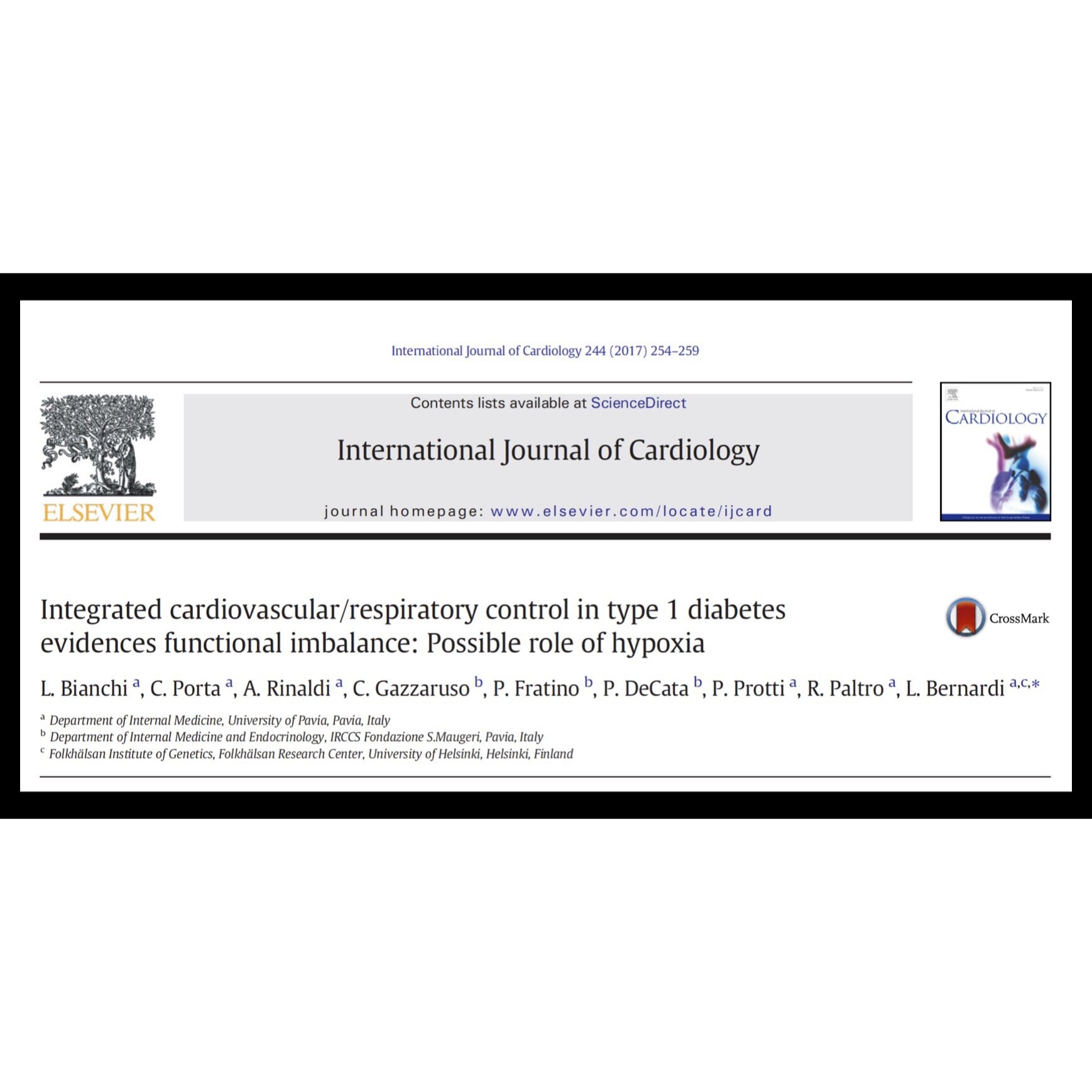
Bianchi L, Porta C, Rinaldi A, Gazzaruso C, Fratino P, DeCata P, Protti P, Paltro R, Bernardi L. Integrated cardiovascular/respiratory control in type 1 diabetes evidences functional imbalance: Possible role of hypoxia. Int J Cardiol. 2017;244:254 – 259.


